Have you ever thought about the secrets hidden just beneath your skin? It might seem like it’s just a simple cover, but each layer has its own special job. The top layer, called the epidermis, works like a shield to block out unwanted germs and irritants. The middle layer, known as the dermis, provides strength and flexibility, almost like a trusty support beam. And deep inside, the hypodermis acts as a natural cushion, insulating and keeping you comfortable. Today, let’s explore how these layers work together every day to keep you safe and why understanding them could change the way you care for your skin.
Comprehensive Overview of the Layers of Skin
Our skin is the largest organ in our body, and it stands as our first line of defense. It blocks out bacteria, protects us from strong UV rays and pollution, and keeps everyday environmental challenges at bay. In a simple way, it works hard to keep our inner organs safe and sound.
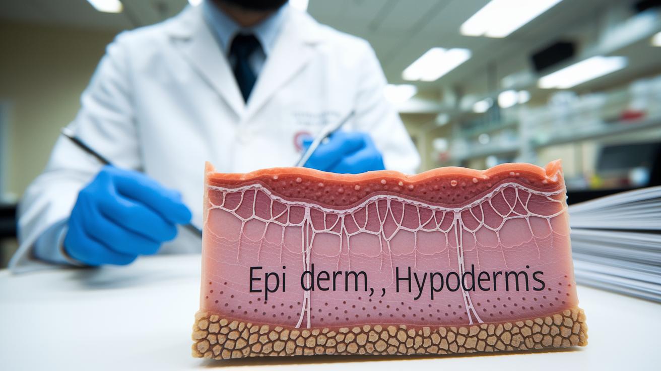
Our skin has three main layers. The outermost layer, the epidermis, is like a visible shield that stops many threats from reaching us. Right under it lies the dermis, full of collagen and elastin fibers that give our skin its strength and elasticity, think of it as the firm, supportive cushion underneath. Deeper still is the hypodermis, made up of fat and connective tissue that not only cushions our muscles and bones but also helps keep our body temperature just right.
This clear breakdown is just a start. Each layer plays its own important role, much like different parts of a well-crafted sandwich. Together, they create a complete system of protection and support that keeps us healthy every day.
Anatomy and Function of the Epidermis Layer in Skin

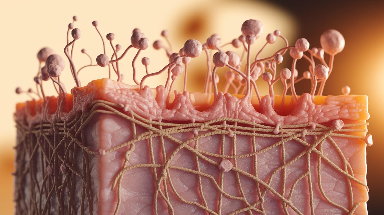
Did you know that an infant's skin can renew itself so quickly that the very first cells get replaced in just a few weeks? The epidermis is the outer layer of the skin, made up of five smaller layers that work together like a team. At the very top, the stratum corneum acts like a tough shield, while at the bottom, the stratum basale is where new skin cells are born. In these layers, cells called keratinocytes produce keratin, a strong protein that helps form a water-resistant barrier. Alongside these cells are melanocytes, which make the pigment that gives your skin its color.
Every 28 to 40 days, your skin gets a complete refresh. It’s much like replacing old bricks in a protective wall. New keratinocytes are made in the stratum basale, then slowly move upward as they mature. By the time they reach the top, they’ve become flat, dead cells that trap moisture inside and keep bacteria and other outside aggressors at bay. This constant renewal keeps your skin healthy, making sure any small damage is quickly fixed while maintaining a strong natural defense.
Structural Features of the Dermis Layer of Skin
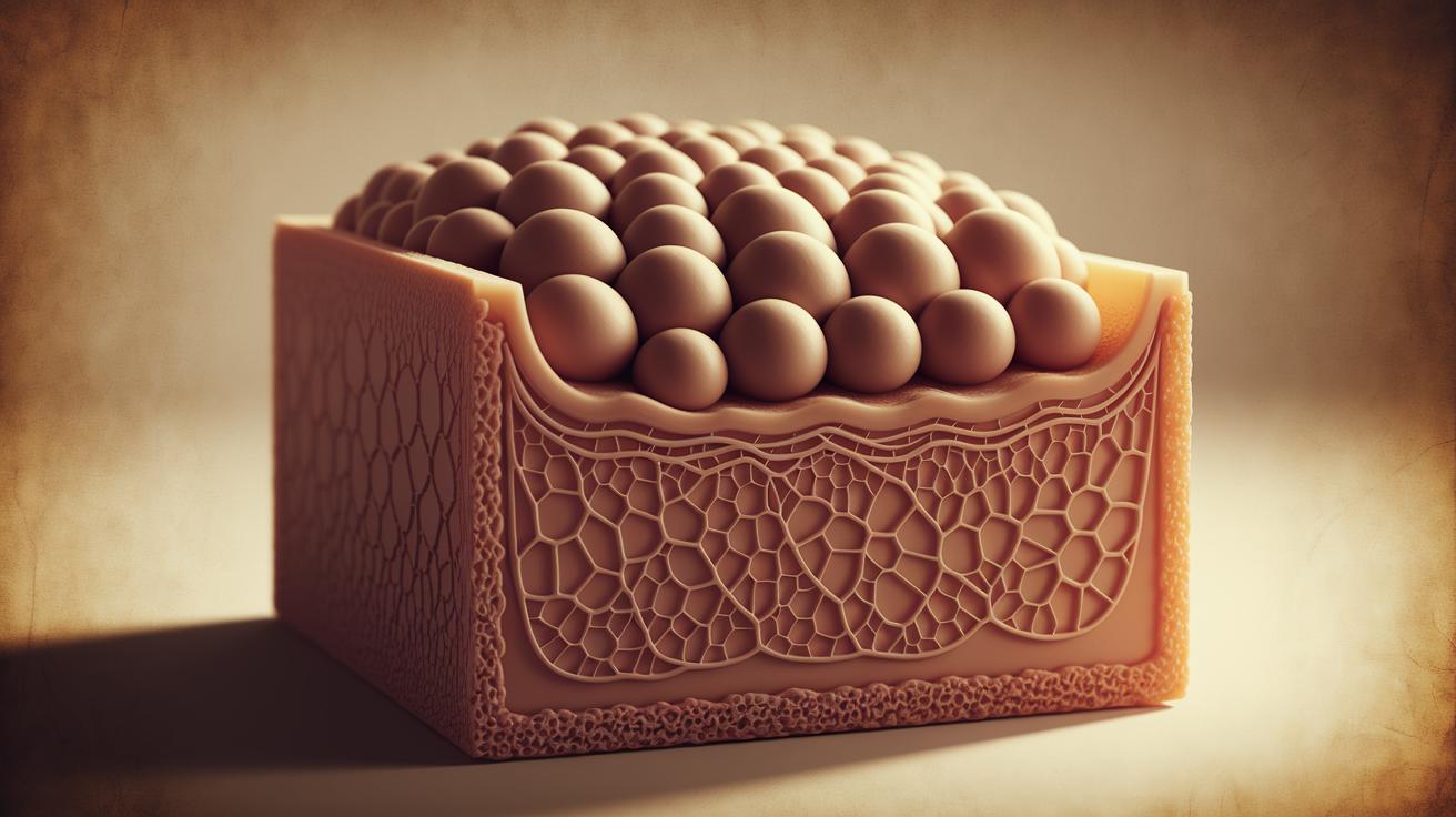
Right below the epidermis, you'll find the dermis, a tough and dependable layer that gives our skin strength and flexibility. The dermis is built from collagen and elastin fibers. Collagen makes the skin strong, while elastin helps it bounce back after stretching.
This layer is also home to key players like blood vessels, sweat and oil glands, hair follicles, and nerve endings. These parts work together to keep your skin nourished, sensitive to touch, and aware of temperature changes.
| Component | Function |
|---|---|
| Collagen fibers | Tensile strength |
| Elastin fibers | Elastic recoil |
| Blood vessels | Nutrient delivery & temperature regulation |
| Sweat glands | Thermoregulation |
| Oil (sebaceous) glands | Lubrication & barrier support |
| Nerve endings | Touch, pain, temperature sensation |
All these fibers and cells do more than keep the skin in place. The collagen and elastin form a sturdy framework, while the blood vessels and glands help your skin stay hydrated and adjust to the heat or cold. Deep nerve endings let you feel what’s happening around you. In short, these features join forces to keep your body’s largest organ healthy and ready for anything.
Composition and Role of the Hypodermis Layer in Skin
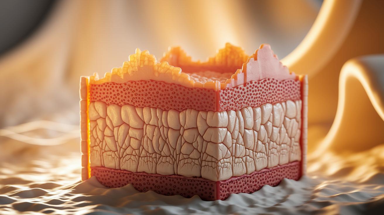
The hypodermis, often called the subcutis, lies just under your skin's surface. It's mostly made of soft, fatty cells and connective fibers that help hold everything together. These fatty cells store energy, while the supportive fibers act like tiny bridges linking your skin to the muscles below, keeping your body stable and running its natural processes.
This layer does more than store energy too. It works as a natural insulator, protecting you from harsh hot and cold temperatures. Plus, it serves as a cushion that absorbs shocks and eases pressure from everyday bumps and knocks, guarding your muscles and bones from harm.
All in all, the hypodermis plays a vital role in keeping your skin healthy. It offers reliable protection, an energy reserve, and steady support to help you face daily life comfortably.
Comparing the Layers of Skin: Anatomy and Interactions
Our skin works like a well-coordinated team. The outer layer, known as the epidermis, acts like a waterproof raincoat. It keeps moisture in and stops harmful substances from seeping in. Just beneath, the dermis steps up with a strong, flexible build and sends gentle signals through nerve endings to your brain. Think of the epidermis as the shield and the dermis as the support that keeps that shield robust.
Then there’s the bond between the dermis and the hypodermis. Here, the strong web of collagen and elastin in the dermis meets a soft layer of fat and connective tissue. The hypodermis not only cushions you from bumps and pressure but also stores energy that aids in healing and regeneration. This snug fit spreads out physical forces smoothly so no single area bears too much pressure.
Together, these three layers form a smart, adaptive system that responds to everyday changes. Whether it’s handling minor bumps, keeping germs at bay, or adjusting to temperature swings, our skin works continuously to protect and renew itself.
Visualizing the Layers of Skin with Diagrams and Imaging Techniques
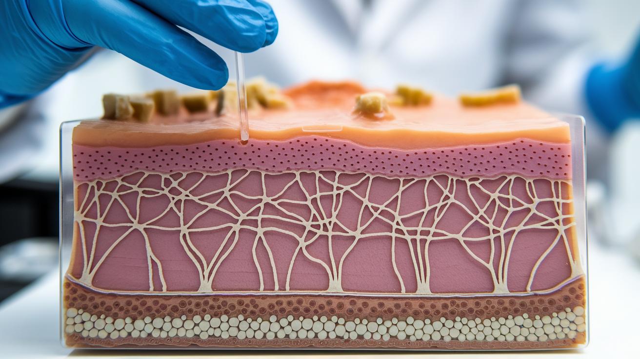
Cross-sectional sketches, histology slides, and other diagrams provide a clear view into how our skin is organized. These images show the outer skin layers, the busy network of collagen and elastin fibers in the middle layer, and the gentle spread of fat cells underneath. Picture it like a simple map where every layer has its own space, making even complex details easy to understand for both patients and professionals.
Advanced imaging methods like confocal and electron microscopy take things a step further by revealing tiny details. They let you see the careful arrangement of skin cells and the structured networks in the deeper layers, almost like zooming in on a vibrant, living landscape. These tools not only confirm the detailed build of our skin but also give us a close-up look at how elements like blood vessels and sweat glands interact, enriching our understanding with real, evidence-backed insights.
Final Words
In the action, we explored the layers of skin by breaking down the epidermis, dermis, and hypodermis. We examined how each layer plays its role in protection, strength, and cushioning while revealing the science behind their interactions.
We also highlighted how visual aids and imaging techniques help illustrate skin layer anatomy in clear terms. Every step reinforces that a better understanding of the layers of skin empowers your cosmetic decisions with genuine, evidence-based insights for a brighter, more confident future.
FAQ
What are the three layers of your skin?
The three layers of your skin include the epidermis as the outer barrier, the dermis for support and sensation, and the hypodermis for cushioning and insulation.
What are the five layers of the epidermis of the skin?
The five layers of the epidermis are the stratum corneum, stratum lucidum (found in thick skin), stratum granulosum, stratum spinosum, and stratum basale, each playing a role in skin renewal.
What does the claim of 7 layers of skin mean?
The idea of 7 layers of skin is a common misunderstanding; medically, skin is categorized into three main layers while only the epidermis is divided into several sublayers.
How are the skin layers shown in diagrams and labeled?
Diagrams typically label the outer epidermis, the middle dermis, and the innermost hypodermis, often highlighting the epidermal sublayers to explain each section’s role.
What are the functions of the different skin layers?
The epidermis protects against pathogens and moisture loss, the dermis offers strength and sensory feedback, and the hypodermis cushions and insulates the body.
What is the Layers of Skin Project?
The Layers of Skin Project is an educational effort designed to illustrate skin anatomy with clear diagrams and labels, helping both patients and professionals understand each layer’s function.

