Have you ever stopped to wonder about the hidden world beneath your skin? Your skin isn’t just a cover; it’s a natural barrier that protects you from everyday challenges. In this guide, we use clear visuals to take you on a simple tour of your skin’s layers. We start with the outer layer that defends you, move on to the middle layer that provides strength, and finish with the inner layer that cushions you. Each part plays its role, working together to keep you healthy and vibrant.
Comprehensive Skin Diagram: Labeled Overview of Cutaneous Structure

The skin is our body's biggest organ and covers about 2 square meters. Its thickness changes a lot, for instance, the gentle skin on your eyelids is around 1.5 mm, while the skin on your palms and soles can be up to 5 mm thick. This amazing organ does many things. It acts like a first line of defense against bumps and harmful environmental factors, helps your body create vitamin D, and uses small chemicals to send important signals.
The skin is built in three main layers, each with its own job. The top layer shields the more delicate tissues underneath, and the deeper layers give your skin strength, flexibility, and a little cushion. For more details in plain language, check out our page on "layers of skin".
• Epidermis
• Dermis
• Hypodermis (subcutaneous tissue)
• Accessory structures (hair, glands, nails)
The skin isn’t just a covering, it’s a tough barrier that protects us from scrapes, chemicals, and pollution. It also helps control water balance and temperature. By blocking too much sun, it helps prevent damage while still letting us benefit from vitamin D. Plus, it produces important chemicals to kick-start healing when needed. This diagram gives you a clear look at how each part of the skin works together to keep you healthy.
Epidermal Diagram: Cross-Section Chart of the Epidermis

Our skin’s outer layer, called the epidermis, is like a natural shield that keeps irritants out and locks moisture in. It’s made up of five layers that work together much like a dependable security system protecting your home.
The very top layer, known as the stratum corneum, is built from dead, flat cells that form a tough barrier. Beneath this, in thicker parts of the skin, lies the stratum lucidum. This layer offers extra strength with a smooth, almost glass-like feel. Right under that is the stratum granulosum. Here, cells packed with small granules help your skin renew itself and hold in moisture. Next comes the stratum spinosum. This layer features cells with tiny spines and creates a supportive structure where immune cells, like Langerhans cells, stay ready to fight off invaders. Finally, at the very base is the stratum basale. This is where new skin cells are born, including melanocytes that add color and Merkel cells that help with touch.
Even though the overall thickness of the epidermis ranges from 0.05 mm to 1.5 mm, every part gets its nourishment through simple diffusion since there are no blood vessels here. This clever design helps the skin keep renewing itself every day.
The differences in how thick each layer is and the balance of these special cells play a big role in how our skin handles injuries, reacts to the environment, and even in how treatments are designed to improve your skin’s barrier and overall health.
Skin Diagram: Radiant Anatomy Visuals

Our skin is built like a layered cake, and one of its main parts is the dermis. This layer has two sections. The upper part, called the papillary layer, is made of soft, loose tissue with tiny blood loops and nerve clusters that help us feel light touches. The lower section, the reticular layer, is packed with strong collagen and stretchy elastin fibers. It usually ranges from 0.3 to 3 mm thick and acts like a supportive base right under the skin’s outer cover.
In the reticular layer, collagen fibers work hard to make your skin strong, while elastin fibers let it stretch and snap back into shape. Tiny blood vessels wind their way through this dense web, delivering nutrients and whisking away waste. Nerve endings are also busy here, sending signals about touch and temperature. Plus, hair follicles along with sweat and oil glands are tucked into this area, helping to keep your skin moisturized and balanced. When you picture these elements together, you see a clear, detailed map of how your skin is put together.
This well-organized structure is what lets your skin stretch, absorb bumps, and stay strong every day, making it a true marvel of natural engineering.
Subcutaneous Diagram: Partition Schematic of the Hypodermis

The hypodermis, or tela subcutanea, is a vital layer under your skin. It works like a cushion to keep you warm and helps absorb everyday bumps.
This layer is mostly made of fat and loose connective tissue, and its thickness can change depending on where it is and who you are. It not only stores energy and keeps your body insulated but also carries larger blood vessels and nerves that reach deeper tissues.
Because it’s soft and flexible, the hypodermis acts as a buffer between your skin and the muscles or bones underneath. This setup helps you move easily and takes some pressure off the skin.
Working closely with the dermis, the hypodermis supplies important blood flow that makes your skin strong and stable. Altogether, this multi-layered system is key for a balanced skin structure by absorbing shocks, regulating your body temperature, and linking the outer layer to the inner parts of your body.
Skin Appendages Diagram: Visual Guide to Hair, Nails, and Glands
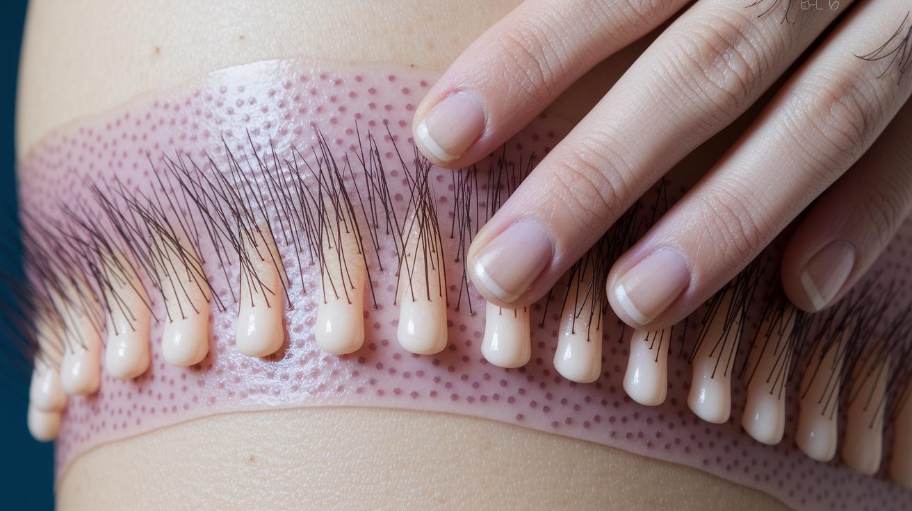
Your skin starts its magic in the dermis, where hair, nails, and various glands are born. This deep layer shows just how every part of your skin works together.
Hair follicles begin their journey here and are quite complex. They work with tiny muscles (called arrector pili) that give you goosebumps and with oil glands that spread natural moisture over your hair. These follicles go through a clear cycle: a growing phase (anagen) when the hair gets long and strong, a shorter phase (catagen), and then a resting phase (telogen) when the old hair falls out, making room for the fresh start.
The nails are another neat part of your skin. Each nail has a hard plate protected by soft folds, a special growth area called the matrix, and a nail bed rich with skin and blood vessels. Together, they shield your fingertips and toes, helping you with everyday activities.
Then there are the sweat glands. Eccrine glands spread across your skin and help cool you down by releasing sweat. Meanwhile, apocrine glands, mostly found in spots like your armpits and near the groin, add to your natural body scent and give a unique touch to your skin’s design.
Skin Diagram of Vascular and Nerve Networks

Our skin is home to a vast network of blood vessels that work like tiny highways. Blood flows from several sources, direct cutaneous, musculocutaneous, and fasciocutaneous, to form two main layers. The top layer, called the papillary layer, and a deeper layer, known as the reticular layer, keep our skin healthy and help control body temperature by spreading heat around.
Deep in the dermis, nerve fibers create a detailed map that connects our body to the world. These fibers include mechanoreceptors, like Meissner’s and Pacinian receptors, which pick up soft touches and vibrations. Other receptors, called nociceptors, alert us if something might hurt, while thermoreceptors help monitor temperature changes. These nerve cells stretch all the way from the inner layers up to where the dermis meets the outer skin, making sure every part of our skin can send signals when something is off. It’s a bit like having a smooth road system that quickly delivers messages, ensuring our skin stays fully aware of what’s happening around us.
This clear picture of where blood vessels and nerves lie is key in treatments that aim to protect or restore the skin’s natural functions.
Annotated Skin Diagram Highlighting Dermatological Conditions

Mapping common skin conditions on this diagram helps you see exactly where the skin is affected. It uses clear markers to show areas troubled by issues like alopecia areata, onychomycosis, and bromhidrosis. For instance, alopecia areata points to spots where the body’s immune system attacks the hair follicles, leading to hair loss. Onychomycosis shows where nail bed and plate infections might occur from fungi, and bromhidrosis marks areas around sweat glands that may cause odor issues.
| Condition | Anatomical Site |
|---|---|
| Alopecia areata | Hair follicles |
| Onychomycosis | Nail bed and plate |
| Bromhidrosis | Apocrine gland regions |
These annotations clearly show how skin conditions change the normal structures of your skin. A marker on the hair follicles can indicate disrupted growth due to an autoimmune reaction. Changes in the nail area point out where the natural barrier might be weakened and vulnerable to fungal infection. And, markings near sweat glands help highlight challenges with sweat regulation. This easy-to-read layout aids both clinicians and patients by pinpointing problem areas and guiding targeted treatment options.
Final Words
In the action of breaking down our skin diagram and its detailed layers, we explored the structure and functions of the epidermis, dermis, hypodermis, and skin appendages. Each section offered a clear look at how these elements work together.
We also highlighted the role of vascular and nerve networks along with common dermatological conditions. The diagram serves as a handy guide to understanding cosmetic treatments and how safe procedures are built on solid science and clear visuals.
FAQ
FAQ
What are 7 layers of skin?
The phrase “7 layers of skin” often refers to a detailed diagram that combines the five strata of the epidermis (corneum, lucidum, granulosum, spinosum, basale) with the dermis and hypodermis.
What are the 5 layers of the skin?
The five layers refer to the epidermis in thicker skin and include the stratum corneum, stratum lucidum, stratum granulosum, stratum spinosum, and stratum basale.
What layer of skin do you feel pain in?
Pain is mainly sensed in the dermis, which houses the majority of the nerve endings responsible for tactile and pain sensations.
Where is the thinnest skin on your body?
The thinnest skin is found on the eyelids, where its minimal thickness allows for delicate flexibility.
What is a skin diagram and how is it used?
A skin diagram is a clearly labeled illustration of the skin’s layers, used to educate about the anatomy and physiology of the skin, making it accessible for both kids and professionals.

